Orthopedic injuries in pets can be particularly severe and painful. If you have ever broken a bone or needed orthopedic surgery, you know that the diagnosis, treatment, and recovery can be intensive. Similar to human medicine, veterinary orthopedic surgery requires a specialist’s expertise, and Stack Veterinary Hospital is fortunate to have one of the few board-certified veterinary surgeons in Onondaga County on our team. Dr. Jan MacDonald has more than 25 years of experience in veterinary orthopedic and soft-tissue surgery, and has been part of the Stack veterinary team since 1992.
We hope your furry friend experiences life-long bone and joint health; however, injuries are sometimes inevitable. Common injuries treated by our orthopedic surgery department include bone fracture reduction and repair, cranial cruciate ligament repair, and luxating patella repair.
Bone fractures in pets
Bone fractures commonly occur in pets who are accidentally dropped or stepped on, or who are hit by a car. Any bone can be fractured, but leg bones are the most common, and they require reduction and stabilization for proper healing. Some fractures can be treated with casts or splints, but many require surgical repair for optimal healing and the pet’s return to full function. Surgical repair includes a variety of devices that keep bones correctly positioned so they will heal, such as:
- Intramedullary (IM) pins — IM pins are long metal rods that can be placed through the center of long bones to keep fracture pieces aligned.
- Bone screws — Small pieces of fractured bone can be held in place with bone screws to allow healing.
- Cerclage wire — Metal wire is sometimes used to encircle fracture fragments and hold them in place.
- Bone plates — Some bone pieces can be held in position with a long metal plate with holes through which screws are placed into the bone.
- External fixators — Severe fractures may require more advanced equipment, such as external fixator devices, which provide stability using several metal rods that are placed perpendicularly through the bone and connected externally to longer rods.
Following fracture stabilization, bones often require several months to regain their original stability and function. Pets are typically able to walk on the affected limb while they are healing, since the surgically implanted devices stabilize the broken bones. Rehabilitation is vital for each pet’s recovery, and can improve her quality of life, enhance range of motion, and reduce pain. Our hospital’s rehabilitation services, led by our certified rehabilitation practitioner, can dramatically improve your pet’s healing.
Cranial cruciate ligament rupture
The cranial cruciate ligament is part of an X-like ligament arrangement in your dog’s knee joint that connects the distal femur to the proximal tibia. The ligament helps provide joint stability by allowing flexion and extension, but resisting abnormal side-to-side and front-to-back motion.
Cranial cruciate ligament rupture, which is the reason for most rear leg lameness in dogs, can occur as an acute injury, or as chronic degeneration. Acute or traumatic injury often occurs when a dog is running, abruptly changes direction, and the knee abnormally rotates. Most of the dog’s body weight is suddenly shifted to the cruciate ligaments, exposing them to excessive rotational and shearing forces, and causing a tear or rupture. Chronic ligament degeneration is a progressive ligament weakening that often begins when the ligament is stretched or partially torn from a mild injury.
Cranial cruciate rupture treatment often includes surgery to reconstruct the knee joint by redistributing forces and eliminating the need for the ligament. Joint stability can also be restored by replacing the damaged ligament with suture-like materials. Pets are typically mobile shortly after cranial cruciate repair surgery, and rehabilitation can enhance healing and reduce pain.
Patellar luxation
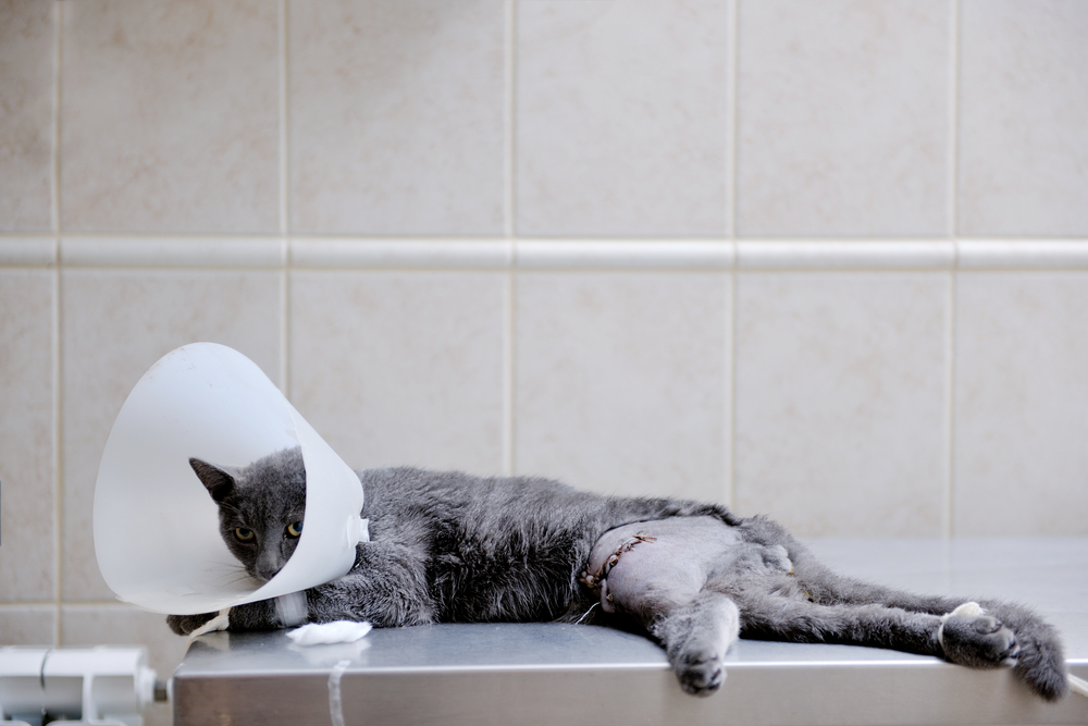
The patella, or kneecap, rests in the patellar groove at the distal femur, and glides up and down in this depression with normal knee movements. Patellar luxation occurs when the developed patellar groove is too shallow to hold the patella in place. During knee flexion, the patella deviates laterally or medially from its normal position. Patellar luxation severity ranges from mild luxation that occurs intermittently only during activity, to permanent dislocation with contracture of associated ligaments and tendons.
Patellar luxation treatment involves surgical deepening of the patellar groove so the patella stays in its normal position during knee movement. The anatomical structures supporting the patella may also be reconstructed to better stabilize the patella in its normal position. After surgery, rehabilitation helps to maintain normal knee joint range of motion during healing and to reduce pain.
Orthopedic procedures in pets are always considered major surgery. They often involve significant pain and a lengthy recovery, and activity must be carefully restricted to allow proper healing and reduce reinjury risk; however, movement is important to prevent muscles and ligaments from weakening. Rehabilitation is a critical component of any orthopedic surgical procedure, as it allows for controlled movements that will help your pet use her muscles, bones, and supporting structures safely to enhance healing and control pain without further injury. Our rehabilitation team will happily help you develop a specialized plan for your pet’s recovery.
If you have questions about orthopedic injuries, or how our hospital’s orthopedic surgery or rehabilitation services can help your pet, contact us.


Leave A Comment